Islet imaging
Pancreatic islets are beautiful islands of endocrine cells that make life-sustaining hormones. We study islets in isolation and in pancreatic tissue to understand how their cellular changes relate to hormone secretion defects in diabetes.
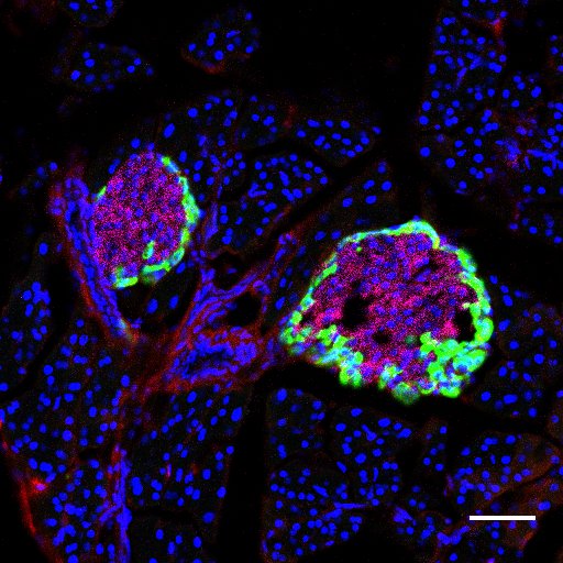
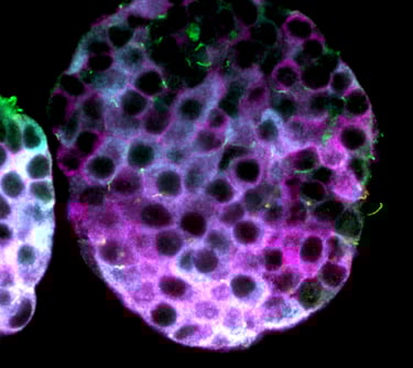
Normal islet
The islets of Langerhans are clusters of endocrine cells located within the pancreas, seen here by glucagon (green) and insulin (magenta) staining amid exocrine tissues.
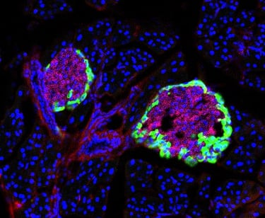
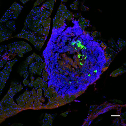
T1D islet
Autoimmune cells infiltrate islets in type 1 diabetes, causing islet cell destruction and loss of insulin secretion, as seen in this diabetic NOD mouse pancreas.

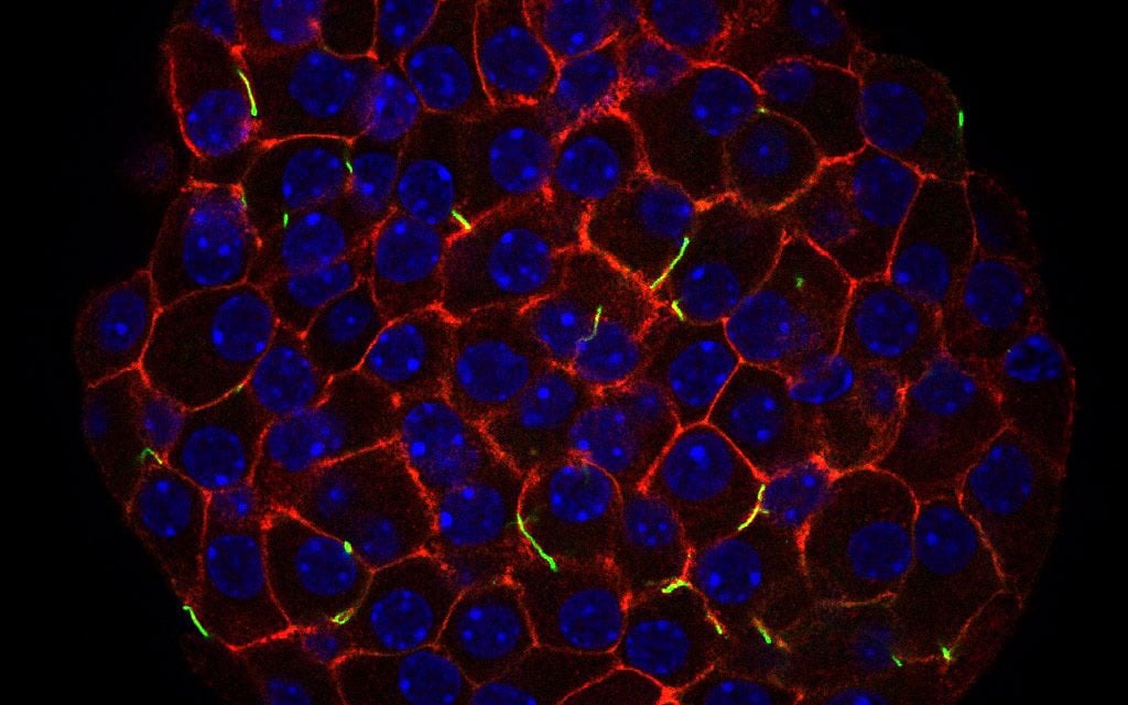
Making islets glow
We transduce and fluorescently label islets to study expression of proteins regulating cilia and islet function.
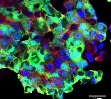

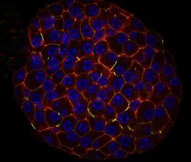
Adenoviral transduction of islets
human islet, ift88 kd (green)
GPCR expression on beta cells
mouse islet, glp1r (magenta)
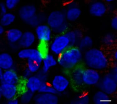
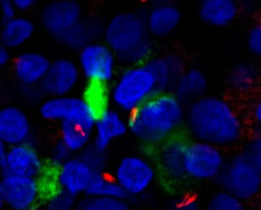
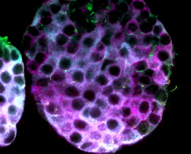
Primary cilia in human islet
Arl13b (red), glucagon (green)
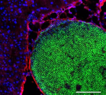
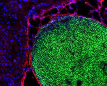
Immune infiltration in T1D
T cells (green) replacing entire islet