Highlight 2: Scanning EM of Islet Primary Cilia

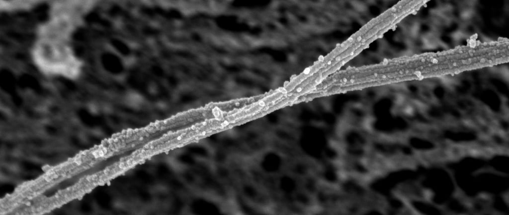
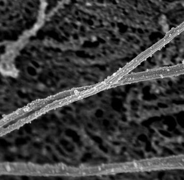
Understanding organelle function starts with seeing what it looks like. Using scanning electron microscopy, we have uncovered a more complex and dynamic structure than previously appreciated. Through innovations in membrane-extraction techniques and advanced instrumentation, we found that the canonical 9+0 microtubule arrangement does not persist along the entire axoneme. Instead, the axoneme exhibits progressive structural evolution, as microtubule doublets transition to singlets toward the tip, and the axoneme adopts a helical twist that has a distinct chirality in mouse versus human tissues. The ciliary tip emerges as a specialized domain with reduced microtubule number and a dense ciliary cap, potentially relevant to its role as a dedicated sensory and signaling hub. These discoveries may help us understand cilia mechanotransduction and the structural basis of motility in pancreatic islets.

6/7/20241 min read

Combining SEM with immuno (yes it is even harder):

Scanning EM of human islet cilia:

Primary Cilia: Sensory Antennas of Pancreatic Islets

Research

Exploring cilia's role in islet cell function.

Science

DISCOVERY

jing.hughes@yale.edu

+1-203-737-2939

© 2025. All rights reserved.